Description
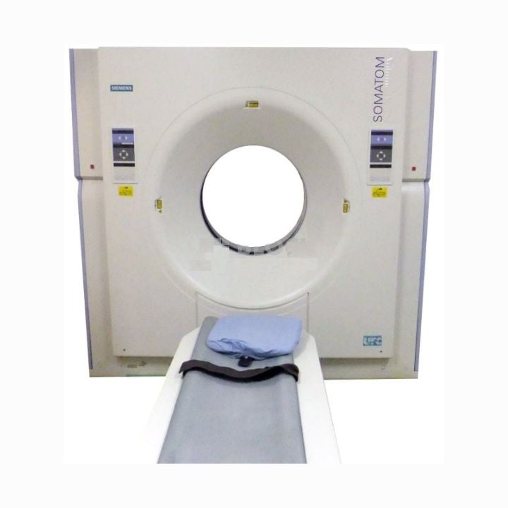
The refurbished Siemens SOMATOM Volume Zoom CT machine is an entry-level 4-slice CT scanner designed to introduce multislice imaging to your clinic at a minimal investment. Our used Siemens SOMATOM Volume Zoom CT scanners are equipped with Siemens’ proven scanning technology, including the CT³ Volume Scanning technique, the Siemens Lightning UFT detector array, the SureView image reconstruction algorithms and more.
Each refurbished Siemens SOMATOM Volume Zoom CT scanner allows for low-dose, thin-slice acquisition with runcompromised reconstruction and streamlined workflow.
The Volume Zoom is an excellent machine for someone new to the CT market and wants to add value to their practice and get a good return on investment. It is a user-friendly machine with great price/performance.
Refurbished Siemens SOMATOM Volume Zoom Features
- 500ms rotation
- 5.3 MHU tube
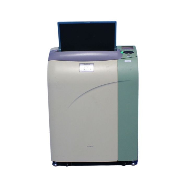
- Syngo user interface
- 60kW generator
- 160 slices in 20-second breath-hold
Spiral Scanning Technique:
- Ultra-fast scanning technique with continuous table feed
- Up to 80 mm/s volume coverage for minimized examination times for live animals
- Data acquisition of an entire anatomical volume up to 157 cm without pause
- Maximum Table Load 200 kg / 450 lbs
- Maximum Gantry Aperture 70 cm.
- Maximum FOV Diameter 55 cm
- Slice Thickness/Collimation 0.5 mm – 10 mm
- Slice Increments 0.1 mm – 10 mm (increment is the distance between the center of adjacent images)
- Scanner Table Pitch 1 – 8. (pitch is the ratio of table feed per rotation to collimation of a single slice)
- Topogram Data:
Min/Max Length: 128/1024 mm
Min/Max Time: 1.6/10.6 sec
Views: Anterior to Posterior (AP), Posterior to Anterior (PA), Lateral (LAT)
Sequence Scanning Technique
- Ultra fast acquisition with or without table feed
- Automatic clustering of scans
| Sequence Slice Width |
| Collimation (mm) |
|
Thickness (mm) |
| 2 x 0.5 |
|
0.5 |
| 4 x 1.0 |
|
1.0 |
| 4 x 2.5 |
|
2.5 |
| 4 x 5.0 |
|
5.0 |
| 2 x 8.0 |
|
8.0 |
| Combi Scan |
| Collimation (mm) |
No. slices fused |
Thickness (mm) |
| 2 x 0.5 |
2 |
1.0 |
| 4 x 1.0 |
2 |
2.0 |
| 4 x 1.0 |
4 |
4.0 |
| 4 x 2.5 |
2 |
5.0 |
| 4 x 5.0 |
2 |
10.0 |
Image Reconstruction:
- 125 ms temporal resolution
- 30 lp/cm spatial resolution
- Scan fields 50 cm; 25 cm (ultra-high resolution collimator)
- Reconstruction Field 5 – 50 cm
- Reconstruction Time 1.5 images/sec
- Matrix 512 x 512 pixels
- Hounsfield Units Scale -1024 to +3071 HU (Extended Scale -10240 to + 40960 HU)
- Tube Current Range 28 – 500 mA
- Tube Voltage 80120, 140 kV