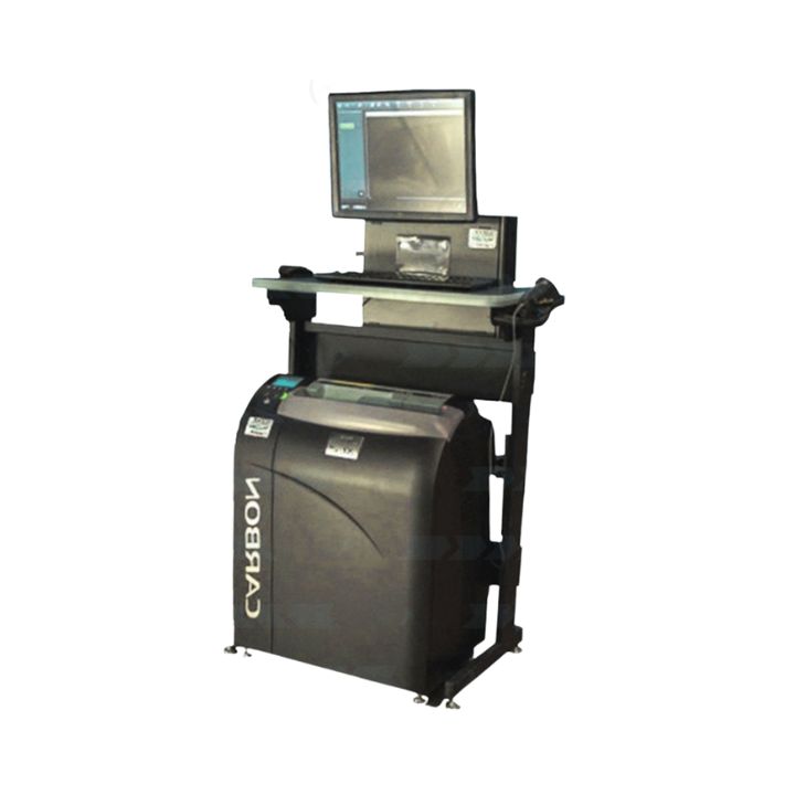
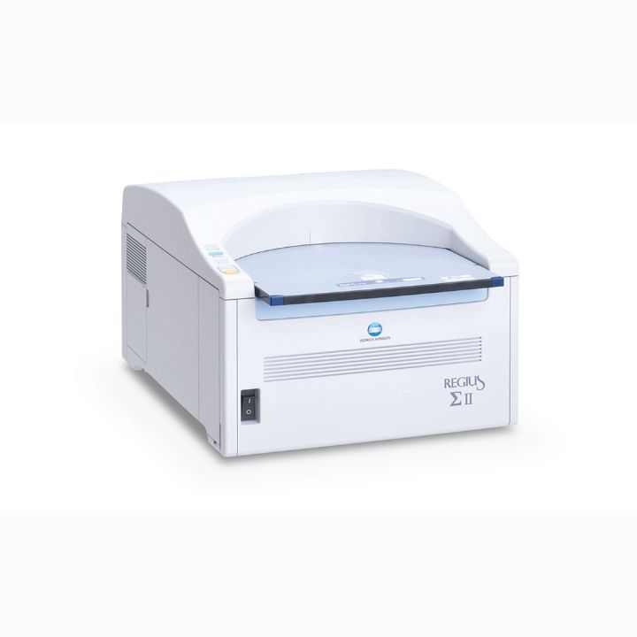
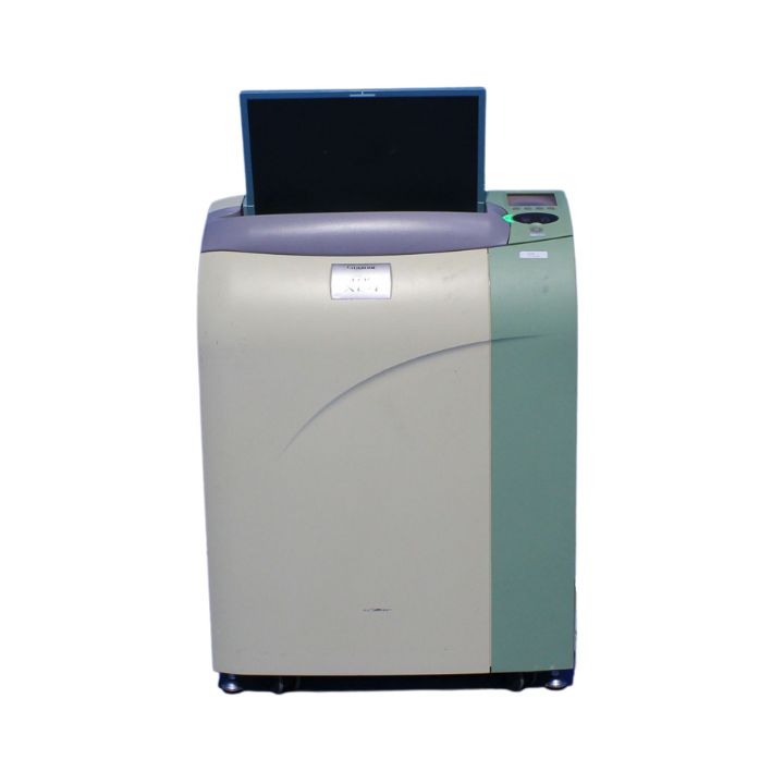
FUJIFILM FCR Carbon XL-2
The FCR Carbon is small and powerful. You get all the productivity and image quality of a full-size CR system in a size that fits in every imaging environment.
The Carbon XL-2 is ideal for distributed applications such as inside the exam room or even a trauma bay with a speed of up to 92 images an hour and image previews in as little as 23 seconds. With that flexibility, the Carbon XL-2 is also a great redundancy solution during peak periods. The XL-2 also features a 50 Reading Mode* for sharp, excellent detail — ideal for extremity exams or anywhere that seeing fine details is critical.
Fast image previews beginning in as little as 10 seconds
Quick throughput – XL-2: 62-92, Fast Scan mode: 87 images/hr 14×17″ X: 43-72 images/hour
100 micron 10 pixels/mm Standard Reading Density
Image Reading 12 bits grayscale, output: 10 or 12 bits Small 2.4 sq. ft. footprint
Specifications:
| Imaging Plates (IPs) and Cassette Sizes | 14″x17″ (35x43cm) 14″x14″ (35x35cm) 10″x12″ 18x24cm 24x30cm 15x30cm The XL-2’s 50 micron capability utilizes Type CH 18×24 & 24x30cm cassettes and single-sided HR imaging plates. |
| Dimensions: | W23.2 x D15.0 x H31.9 inches |
| Weight | 216 lbs. |
| Power Requirements | 110 VAC (5A max) |
*Not intended for mammography use. However, it can be used for specimens.
Accessories:
Additional Flash IIP consoles, advanced imaging capabilities (such as DVD storage, Stitching and more), workstation/reader stand and seismic & mobile laptop IIP and vehicle mounting hardware are available separately.
FDX CONSOLE
PRODUCT CODE: FDXCONSOLE
The FDX Console is the heart of every FDR and FCR system. It represents the culmination of Fujifilm’s extensive experience in image and information processing. The sophisticated interface is intuitive and customizable, and enables fast, easy exam completion and image optimization
New features include:
- Dynamic Visualization™ is Fujifilm’s advanced image processing technology, providing outstanding first-up detail and virtually eliminating the need for post processing.
- Complete common workflow steps in as few as 2-3 mouse clicks
- Worklist views with status icons and thumbnail images
- Virtual Grid™ (option) – improved image quality for images acquired without a grid
- Double click full-screen zoom
- PICC line/edge enhancement toggle
- Dose tracking and management tools
- Optional 2MP secondary monitor for PACS comparable preview
Dose optimization features
- Customizable menus help simplify dose-saving techniques.
- Exposure Index (EI) and Deviation Index (DI) guides and tracks deviation from preferred exposure conditions for each exam.
- EI and DI values can be sent in DICOM headers, allowing low dose initiative monitoring.
See more, click less
- Multi-Accession opens multiple studies with separate accessions for the same patient in one acquisition screen
- Auto trimming simplifies offset imaging by automatically detecting the exposure area. The true sized cropped image is sent to PACS, for optimal image display size at both workstations.
- Region of Interest (ROI) image adjustment — unique 2-point ROI function that reprocesses images instantly based on user-selected points of interest.
- Image Stitching (optional) for DR and CR automatically combines multiple adjoining images into a single long length image for scoliosis and long leg studies.
Accessory
Wall mounting hardware for FDX Console
Delivering a refined image processing experience
Dynamic Visualization™ processing tools produce high quality images that aid diagnosis and boost productivity. Optimal first-up display virtually eliminates any need for post processing image adjustments, providing exceptional image quality automatically. Wide dynamic range adaptability and breadth of exam menus help to reduce retakes.
Fujifilm’s Dynamic Visualization image processing automatically recognizes the region of interest and applies the optimum image processing parameters throughout the entire exposure field producing exceptional images with higher window and leveling content for faster, more accurate diagnosis. Additional advanced functions include:
- Dynamic Visualization™ II (upgrade option): Our latest evolution in image processing. Dynamic Visualization II advanced processing with auto recognition of bone, anatomy characteristics, and orthopedic hardware. This image processing software intelligently adapts image contrast and density, based on image, thickness and structural recognition. It improves uniformity in both dense and thin regions for challenging images, for large anatomy and patients, or any low dose or low penetration exams.
- Virtual Grid (option): Processing intelligently simulates grid use, eliminating scatter effect, to improve contrast and clarity for images acquired without a grid. Useful in portable exams: simplifies acquisition and positioning, and eliminates artifacts associated with physical grid misalignment and improper SID.
- Intuitively recognizes scatter effect in the image
- Precisely tunes contrast and noise control
- Eliminates misalignment factors
- Customizable grid characteristics, grid lines, density and interspacing material

