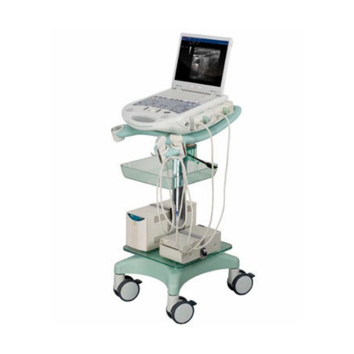
Toshiba Aplio XG Color Doplor Ultrasound
Intelligently designed for precise diagnostic imaging and optimized clinical workflow, the Toshiba Aplio XG ultrasound is considered as today’s most innovative solution that meets the meticulous demand of the medical arena for imaging quality and limitless productivity.
The Aplio XG is engineered with cutting-edge MicroFlow contrast precision imaging mechanism that allows for unparalleled, ultra-clear, high-resolution 4D ultrasonic images featuring the intricate details of the patient’s diagnosed system with clarity. Most preferred by diagnostic mammogram specialists, the Aplio XG is also integrally built with advanced MicroPure clinical technology that helps enable the physician to accurately detect microcalcifications – the biomarker for breast tumor malignancy.
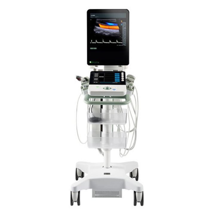
Meet your challenging clinical demands with outstanding performance and comfort.
XarioPrime‘s powerful system architecture, whose intelligent components work and communicate autonomously, supports the most advanced imaging functions. XarioPrime is also easy to upgrade, to keep you abreast of new ultrasound techniques. Excellent diagnostic performance. Operator comfort that inspires productivity. And outstanding connectivity features XarioPrime. Quite simply, the prime ultrasound system for a wide range of clinical applications.
Performance
Gain greater diagnostic confidence with advanced, clinically proven technologies
- XBT Transducer Technology
- Pulse Subtraction THI
- Precision Imaging – Real Time Enhancement of Ultrasound Images
- ApliPure
- Advanced Dynamic Flow
- Low MI Pulse Subtraction
- Trapezoid Imaging
- Panoramic View
- 3D/4D Imaging
Precision Imaging*
(Unique Feature of Toshiba)
- New method of signal processing powered by Intelligent Component Architecture
- Enhance ultrasound beam data by including information from adjacent lines
- Allow early identification of diffuse random noise and structural boundaries
- Improves resolution, more homogenous image, less noise